Auteur/autrice : admin

  • Navigating the challenges of narrow lumbar canal

    Navigating the challenges of narrow lumbar canal

    Dealing with a narrow lumbar canal can be a significant health concern, often leading to discomfort and reduced mobility. It’s crucial to understand this condition, its symptoms, and the available treatment options. This guide provides an insightful overview of narrow lumbar canal, also known as lumbar stenosis, helping you navigate the diagnosis and treatment journey effectively.

    Understanding narrow lumbar canal

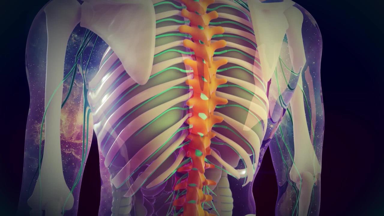
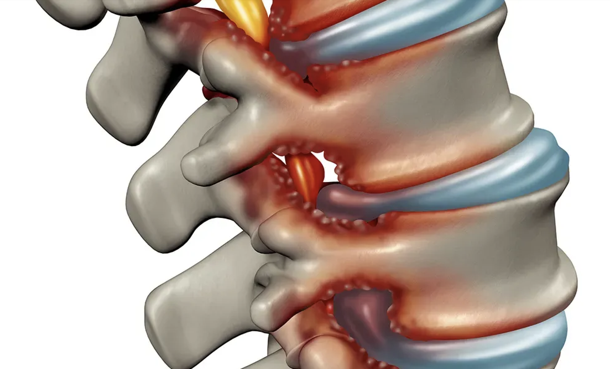
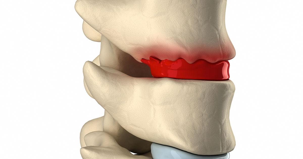
    Lumbar stenosis, commonly known as a narrow lumbar canal, is a degenerative condition resulting from the reduction in the diameter of the lumbar canal. This leads to the compression of nerve roots located within it. Here’s what you need to know about this disorder.

    Diagnosing narrow lumbar canal: how to spot it?

    The narrowing of the lumbar canal is characterized by bilateral pain in the lumbar region and the lower limbs, mainly when the patient is standing or walking. In rare cases, paralysis of the lower limbs and sphincter functions can occur.

    However, symptoms can be subtle and may be confused with other conditions. To confirm the diagnosis, your doctor may recommend imaging tests, such as:

    • An X-ray to identify indirect signs;
    • A CT scan, and especially an MRI, to visualize the canal and its contents;
    • An ultrasound of the lower limb arteries to rule out other causes of lower limb pain.

    Treatments for narrow lumbar canal: what are the options?

    Except in cases of motor deficit, unbearable pain, or paralysis of sphincter functions, a conservative medical approach can be considered with:

    • Pain relievers;
    • Anti-inflammatories;
    • Canal infiltration;
    • Rehabilitation, among others.
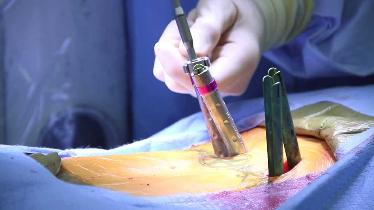
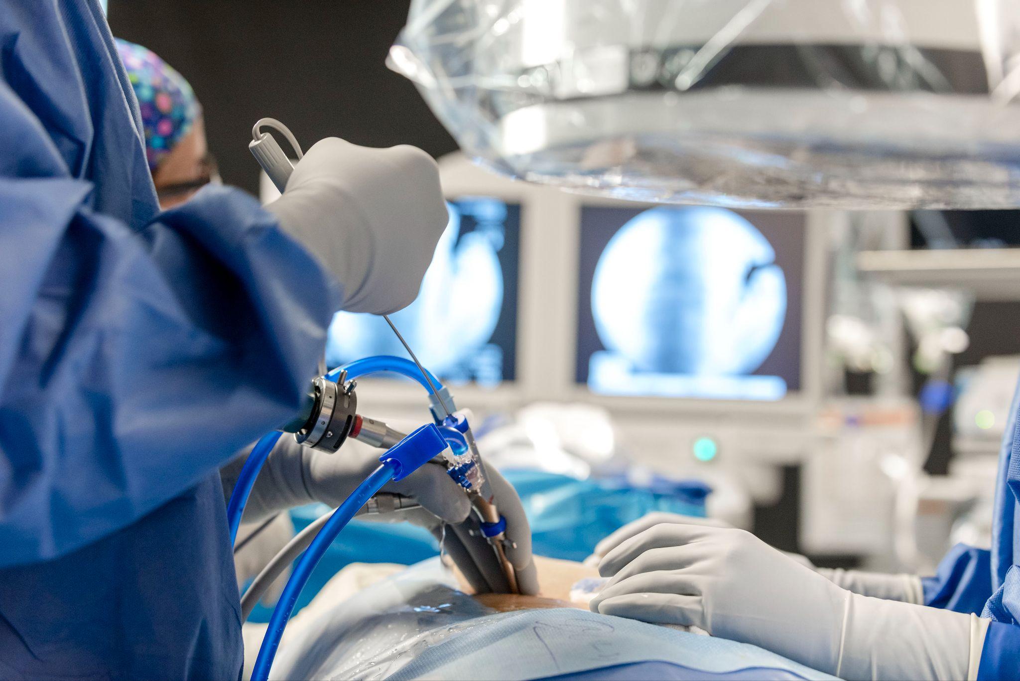
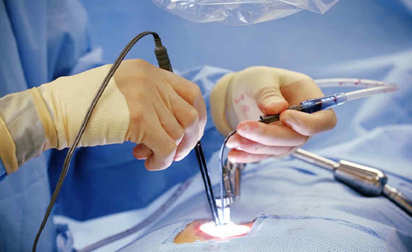
    • If these methods do not provide relief, surgical intervention may be considered to enlarge the canal. This surgery, performed under anesthesia, primarily aims to decompress the nerve elements by removing tissues, bone fragments, or herniated discs obstructing the passage. The surgical technique will depend on the type of stenosis and the elements to be removed.

    Postoperative results: what to expect after narrow lumbar canal surgery?

    Although surgery may not always eliminate all symptoms, especially if the nerve root has been damaged over a long period, most patients experience a significant improvement.

    In general, partial autonomy is regained 24 hours after the procedure, and symptoms such as pain, numbness, and tingling tend to disappear within a few weeks.


  • Causes of lumbar spinal stenosis: A complete guide

    Causes of lumbar spinal stenosis: A complete guide

    The lumbar spinal canal plays a vital role in our spinal health and nervous system function. However, it can sometimes narrow, leading to symptoms of discomfort and pain. This article delves into the underlying causes of lumbar spinal stenosis, offering an expert overview of factors to consider and available solutions for regaining well-being.

    Understanding lumbar spinal stenosis

    The lumbar spinal canal, which houses the spinal cord and nerve roots, can become narrow for various reasons. This narrowing can put pressure on nerve structures, causing a range of uncomfortable symptoms. Understanding the underlying causes is crucial for taking appropriate measures and finding suitable solutions.

    Factors contributing to lumbar spinal stenosis

    • Osteoarthrosis: Osteoarthrosis is one of the primary causes of lumbar spinal stenosis. Degenerative changes associated with osteoarthritis can lead to abnormal bone and tissue growth, reducing space in the canal.
    • Disc herniations: The intervertebral discs located between the vertebrae can weaken over time and compress the lumbar canal. Disc herniations can also cause inflammation contributing to narrowing.
    • Excessive bone growth: In some cases, excessive bone growth, known as osteophytes, can develop in the lumbar canal. This may be a response to previous damage or osteoarthritis.
    • Trauma: Previous injuries, such as fractures or trauma, can contribute to lumbar spinal stenosis by altering bone structure.
    • Genetic factors: Some individuals may be genetically predisposed to developing lumbar stenosis due to their spinal structure.

    The importance of medical consultation

    If you experience symptoms like lower back pain, numbness, tingling, or difficulty walking, consulting a qualified health professional is essential. A specialist in endoscopic spine surgery can assess your situation and recommend the best approaches for treating lumbar spinal stenosis.

    Tailored solutions with endoscopic spine surgery

    If conservative treatments fail to alleviate your symptoms, surgery may be considered. Endoscopic spine surgery, as practiced at specialized centers, offers a modern and minimally invasive option for treating lumbar spinal stenosis.

    Benefits of endoscopic surgery

    • Reduced postoperative pain: Compared to traditional surgery, endoscopic spine surgery is generally associated with less postoperative pain.
    • Quicker recovery: Smaller incisions used in endoscopic surgery allow for faster recovery and a quicker return to daily activities.
    • Less impact on surrounding tissues: The less invasive nature of endoscopic surgery reduces muscle trauma and limits scarring.

    Taking steps for a healthy future

    Lumbar spinal stenosis doesn’t have to compromise your quality of life. Teams at specialized spine surgery centers are committed to providing high-quality care to help you regain comfort and mobility.

    If you suffer from symptoms of lumbar spinal stenosis, consulting an experienced health professional is crucial. Specialized centers are ready to assess your situation and guide you toward the best treatment solutions.


  • Diagnosing Narrow Lumbar Canal: A comprehensive guide

    Diagnosing Narrow Lumbar Canal: A comprehensive guide

    At the Center for Endoscopic Spine Surgery, Dr. Léglise provides insights on diagnosing a narrow lumbar canal.

    Lower back and leg pain can be debilitating, impacting your quality of life and daily activities. If you’re experiencing fatigue, tingling, or cramps in your lower limbs, understanding how to diagnose a narrow lumbar canal is crucial. Dr. Léglise and his team at the Center for Endoscopic Spine Surgery are here to guide you through this essential diagnostic process.

    The structure of the spine

    The spine, also known as the vertebral column, is a marvel of biological engineering. Comprising 24 stacked vertebrae connected by essential joints, it houses the spinal canal – a central opening in each vertebra through which the spinal cord and nerves responsible for sensations and movements in the body below the head travel. The three main joints – the intervertebral disc and the two posterior joints – work in harmony to ensure smooth mobility while providing solid support.

    Understanding narrow lumbar canal

    Narrow lumbar canal, a common condition, typically develops gradually due to spinal osteoarthritis, the natural wear and tear of joints. This osteoarthritis can lead to a narrowing of the spinal canal where essential nerves to the lower limbs and sphincters pass. Picture it like the fingers of an aging hand, which tend to thicken due to osteoarthritis, thus reducing space for nerves.

    Contributing factors and symptoms

    Other factors, like a vertebra slipping (spondylolisthesis), can also cause canal narrowing, though this is less common. When nerves are compressed, symptoms emerge. You might experience limping while walking, a feeling of fatigue, or tingling in your legs after a certain distance.

    Listening to your body: Recognizing the symptoms

    Pain is often the first alert of a narrow lumbar canal. Cramps and a feeling of fatigue in the lower limbs during walking are common. You might even feel as if your legs could give way. If pain forces you to frequently stop walking, you might unconsciously adopt the « caddy » posture. This instinctive forward-leaning gesture helps to artificially open the lumbar canal and foramina, providing temporary relief.

    Accurate diagnosis

    Dr. Léglise’s team at the Center for Endoscopic Spine Surgery conducts a series of comprehensive examinations for an accurate diagnosis. These include:

    • Static and dynamic lumbar radiographs: These images confirm the presence of spinal osteoarthritis and reveal the stability or instability of the vertebrae.
    • CT scan: Particularly effective in visualizing bony aspects, it confirms the diagnosis and specifies the number of affected vertebral levels.
    • MRI (Magnetic Resonance Imaging): The MRI is the examination of choice. In addition to the information provided by the CT scan, MRI shows direct narrowing on the nerves and identifies contributions from both bony structures and soft parts.

    Dr. Léglise’s expertise

    Dr. Léglise and his team are renowned specialists in spinal conditions. The Center for Endoscopic Spine Surgery offers cutting-edge solutions to treat narrow lumbar canal and improve your quality of life. If you’re experiencing symptoms similar to those described here, consult Dr. Léglise and his team for a thorough diagnosis and tailored care for your condition.


  • Managing Degenerative Disc Disease

    Managing Degenerative Disc Disease

    Degenerative lumbar disc disease is a common cause of back pain. Understanding its mechanisms and treatment options is crucial for effectively alleviating these pains and improving quality of life.

    What is degenerative lumbar disc disease?

    Degenerative lumbar disc disease is characterized by the progressive degeneration of intervertebral discs, mainly in the lumbar region. Over time, these discs lose hydration and elasticity, leading to pain often described as a « bar » in the back, which can radiate to other body parts. The L5-S1 discs are most commonly affected.

    Diagnosis and risk factors

    Diagnosing degenerative lumbar disc disease typically involves a clinical examination followed by radiographs and an MRI. Risk factors include natural aging, overweight, certain back pathologies, poor lifestyle habits, lack of physical activity, and genetic predispositions.

    Conservative treatment: The first step

    Medication

    For pain relief during a flare-up, analgesics, anti-inflammatories, and muscle relaxants are often prescribed. These medications help manage symptoms and improve patient comfort.

    Physiotherapy

    Physiotherapy plays a crucial role, aiming to relax muscles, particularly in painful positions, and work on postural realignment. Exercises to strengthen the core and abdominal belt can be incorporated to further support the spine.

    Corticosteroid injections

    In some cases, corticosteroid injections may be necessary to reduce inflammation and pain.

    Back surgery: when other treatments are insufficient

    If conservative treatment fails to sufficiently ease the pain or if the condition becomes too debilitating, back surgery may be considered. The main goal is to decrease lumbar pain by limiting the movements of affected discs.

    Spinal fusion

    Spinal fusion, a surgical procedure where vertebrae are fused together, is an option to stabilize the spine and alleviate pain. This intervention can be performed using modern techniques like mini invasive surgery, coupled with rapid recovery after surgery (RRAC).

    The treatment of degenerative lumbar disc disease starts with conservative approaches like medication and physiotherapy. If these fail, surgical options such as spinal fusion may be considered.

    At the Center for Endoscopic Spine Surgery, Dr. Léglise and his team utilize the latest techniques to ensure optimal care and a quick recovery. If you are suffering from degenerative disc disease, do not hesitate to schedule a specialized consultation.


  • Professionnal sportive could be operated by minimal invasive technique

    Professionnal sportive could be operated by minimal invasive technique

    FC Barcelona’s goalkeeper Marc-André ter Stegen recently underwent a successful back surgery, a pivotal moment in his sports career. This procedure was carried out by Dr. Amélie Léglise, a leading figure in the field of endoscopic back surgery.

    Marc-André ter Stegen’s surgical procedure

    Marc-André ter Stegen, who had been off the field since November 12 due to back pain, underwent surgery on December 8 at a specialized clinic. The operation was performed by Dr. Amélie Léglise, under the supervision of the Club’s Medical Service, at the Bordeaux Mérignac Sports Clinic.

    Dr. Léglise’s expertise in endoscopic surgery

    Dr. Léglise’s specialty, endoscopic back surgery, is a minimally invasive technique effectively treating various spinal pathologies, including those encountered by top athletes like Ter Stegen. This method offers several benefits, including reduced risk of complications and faster recovery time.

    Ter Stegen’s return to the field

    While FC Barcelona remains discreet about the exact duration of Ter Stegen’s unavailability, hopes are high for a swift return, potentially for the Champions League round of 16 in February. The success of the operation and Dr. Léglise’s reputation in endoscopic surgery suggest a positive prognosis.

    Endoscopic surgery: a revolution in treating lumbar pathologies

    Endoscopic surgery, with its precision and less traumatic approach compared to traditional surgical methods, represents a significant advancement in treating spinal disorders. High-level athletes, like Marc-André ter Stegen, particularly benefit from this technology, allowing for a quicker return to optimal performance levels.

    If you are experiencing back pain or wish to learn more about treatment options, do not hesitate to schedule an appointment with Dr. Léglise at the Center for Endoscopic Spine Surgery in Bordeaux to benefit from her expertise.


  • Minimally invasive lumbar in France

    Minimally invasive lumbar fusion at the Center for Endoscopic Spine Surgery in Bordeaux, France

    The health of the spine is essential for overall well-being. Among the procedures designed to restore spinal stability and functionality, minimally invasive anterior lumbar fusion stands out for its advanced technique.

    At the Center for Endoscopic Spine Surgery, we highlight this procedure for its numerous benefits.

    What is minimally invasive lumbar fusion?

    Minimally invasive lumbar fusion involves fusing two adjacent vertebrae to stabilize a region of the spine. A device known as a « cage, » which is essentially a spacer, is placed between these vertebrae after being filled with graft material, either synthetic or harvested from the patient’s pelvis. Often, an additional plate is used to enhance stabilization.

    How is the procedure performed?

    Carried out under general anesthesia, the operation usually lasts between 1 and 2 hours. The surgeon accesses the spine through an anterior incision, either in the center or slightly off-center of the lower abdomen.

    What are the associated risks?

    Like any surgery, minimally invasive lumbar fusion carries certain risks, although they are rare. These risks include hemorrhagic complications, temporary neurological complications, mechanical problems related to the implant, and infection risk.

    What to expect after the operation?

    Our patients can typically get up the evening or the day after the surgery with the help of our dedicated physiotherapists. Returning home is usually considered as soon as intestinal transit resumes, generally between the 2nd and 4th postoperative days.

    Success rate of the procedure

    According to several studies, minimally invasive anterior lumbar fusion has an impressive success rate of 80-85%. We ensure that each patient receives postoperative follow-up to monitor their clinical recovery and bone consolidation, which typically takes 6 to 12 months.

    Minimally invasive lumbar fusion is an advanced surgical option for patients seeking a lasting solution to spinal problems. At the Center for Endoscopic Spine Surgery, we are proud to offer this cutting-edge intervention and support our patients throughout their healing journey.

    Consider consulting with the Center for Endoscopic Spine Surgery in Bordeaux to explore the benefits of this technique.


  • Endoscopic spine surgery : Cervical disc herniation treatment

    Endoscopic spine surgery : Cervical disc herniation treatment

    Cervical disc herniations can be a significant source of pain and discomfort for many patients, profoundly impacting their quality of life by limiting mobility and causing severe pain. However, with recent advances in surgical techniques, an innovative and less invasive approach is now available: endoscopic spine surgery.

    Understanding cervical disc herniation

    A disc herniation occurs when the nucleus of an intervertebral disc escapes through the surrounding fibrous ring. This protruding fragment can press on nerve roots, causing pain radiating into the arm. In more severe cases, the spinal cord itself may be affected, leading to complex neurological symptoms.

    The importance of diagnosis

    In addition to physical examination, the diagnosis of a cervical disc herniation relies heavily on MRI, sometimes supplemented by electromyography or CT scans. Symptoms vary depending on the exact location of the herniation. For instance, a C7 – D1 herniation might lead to weakness and tingling in the fingers, while a C4 – C5 herniation could present as shoulder pain or weakness in the upper arm.

    Endoscopic intervention: A significant advancement

    The goal of cervical disc herniation surgery is to remove the herniation and decompress the nerve. This procedure, performed through a short incision on the anterior aspect of the spine, utilizes the endoscopic system to largely preserve the intervertebral disc. This less invasive approach offers numerous advantages over traditional methods.

    Benefits of the endoscopic technique

    Beyond being less invasive, endoscopic spine surgery results in particularly straightforward postoperative recovery. Patients experience immediate relief and can quickly resume their daily activities. This rapid recovery is a crucial aspect for many patients eager to regain their mobility and quality of life as soon as possible.

    The patient journey: From preparation to recovery

    Prior to the procedure, patients receive attentive and professional care. An orderly escorts them to the operating room, where they are greeted by the anesthetist and their nurse in the pre-anesthesia room. Once sedated, patients benefit from the expertise of the surgical team for about an hour. After surgery, patients are monitored in the recovery room before returning to their ward.

    Rapid and guided recovery

    Recovery after endoscopic spine surgery is often remarkably swift. Upon returning to their room, patients are offered a snack or meal. The IV is removed, allowing them to shower and stretch their legs. Physiotherapists, in collaboration with the surgical team, visit the patient the same evening in their room, and the next morning for the first rehabilitation session.

    A new era for cervical disc herniation treatment

    Endoscopic spine surgery, as performed by our team, represents a significant advancement in the treatment of cervical disc herniations. This less invasive approach provides patients with a faster, less painful recovery while preserving the integrity of the intervertebral disc as much as possible.

    If you are suffering from pain related to a cervical disc herniation, consider consulting with the Center for Endoscopic Spine Surgery in Bordeaux to explore the benefits of this technique.

  • Endoscopic spine surgery: minimizing risks and enhancing recovery

    Endoscopic spine surgery: minimizing risks and enhancing recovery

    Endoscopic spine surgery marks a pivotal advancement in spinal healthcare, revolutionizing the way spinal conditions are treated. This state-of-the-art approach significantly diminishes the risks typically associated with traditional spinal surgeries while expediting the patient’s recovery journey.

    Understanding endoscopic back surgery

    Endoscopic spine surgery incorporates the use of specialized cameras and instruments, introduced through minimal incisions, often just a few centimeters long. This technique stands in contrast to conventional surgery methods, offering a less invasive option. By reducing muscle and bone trauma, it significantly streamlines the patient’s rehabilitation process.

    Ideal candidates for endoscopic surgical procedures

    This form of surgery is particularly beneficial for conditions that don’t require extensive hardware implantation, such as screws or rods. It’s proven effective for managing sciatica, cruralgia, narrow lumbar canals, and various forms of disc herniations and lumbar stenosis. A notable advantage of this method is its potential to reduce the need for more invasive lumbar fusion surgeries, therefore avoiding extensive vertebral fusions.

    Advantages of endoscopic spine surgery

    Choosing endoscopic spine surgery translates into numerous patient benefits:

    • Accelerated recovery timeline, enabling patients to return to their daily activities sooner.
    • Smaller incisions lead to reduced postoperative discomfort and lower dependency on pain medication.
    • Significant decrease in blood loss during surgery.
    • Lower risk of infection due to the minimally invasive nature of the procedure.
    • Preservation of spinal stability by avoiding extensive lumbar fusions.

    The lumbar spine endoscopy procedure explained

    During a lumbar spine endoscopy, patients are placed under general anesthesia for optimal immobility and safety. Through advanced radiological guidance, surgeons meticulously perform the procedure, introducing the endoscopic camera directly to the affected vertebral area. This approach allows for precise nerve decompression, akin to conventional open surgery but with minimized muscle disruption. Post-surgery, careful monitoring of the incision sites ensures proper healing.

    Understanding the risks of endoscopic back surgery

    While endoscopic spine surgery is relatively low-risk, it’s important for patients to be aware of potential complications. Nerve damage, although rare, is a consideration. Infections are also possible but occur less frequently compared to traditional surgery methods. Dural tears, involving the membrane around the nerves, can happen but are a manageable aspect of this surgery.

    Endoscopic spine surgery at our center not only offers a safer, less invasive treatment option but also a quicker path to recovery, reflecting our commitment to advanced spinal care and patient wellbeing.


  • Introducing the Center for Endoscopic Spine Surgery in Bordeaux

    Introducing the Center for Endoscopic Spine Surgery in Bordeaux

    Facing back problems can be daunting, and choosing the right surgical intervention is crucial. This is where the center for endoscopic spine surgery in Bordeaux excels. Located in the heart of Mérignac, this state-of-the-art clinic specializes in a groundbreaking approach for a variety of spinal issues. Let’s delve into why this center, under the leadership of renowned experts, is considered a premier destination for back surgery in Bordeaux.

    Our team of renowned specialists

    At the heart of our center is a dedicated team of medical professionals, each bringing a wealth of expertise to provide top-tier spinal care.

    Dr. Jean Destandau: a neurosurgery innovator

    Dr. Jean Destandau, a University of Bordeaux alumnus, is a trailblazer in neurosurgery. He revolutionized spinal surgery in 1993 with his development of the endoscopic technique, known as endospine. With a successful track record of over 7,000 lumbar disc herniation surgeries, Dr. Destandau is a revered figure in the field.

    Dr. Amélie Léglise: orthopedic surgery specialist

    Dr. Amélie Léglise, renowned for her work in orthopedic and trauma surgery, enhances the center’s treatment spectrum. Her proficiency in minimally invasive endoscopic surgery, especially for lumbar and cervical disc herniations and narrow lumbar canal, is exceptional.

    Dr. Marjory Rué: neurosurgery expert

    Dr. Marjory Rué brings her extensive neurosurgery experience and tenure as a former intern and head of clinic at Bordeaux hospitals to the team. Collaborating closely with Dr. Destandau, she specializes in minimally invasive endoscopic solutions for lumbar and cervical disc herniation and narrow lumbar canal.

    Innovative approach : endoscopic spine surgery

    Endoscopic spine surgery is a cutting-edge advancement in spinal condition treatments. Compared to traditional methods, it offers significant benefits:

    • Tailored to conditions like lumbar and cervical disc hernias, narrow lumbar canal, and spinal synovial cysts.
    • Conducted through a small incision, using a mini-camera to provide a detailed view of the operative area.
    • Benefits include quicker recovery, minimal scarring, reduced muscle trauma, enhanced surgical visibility, lower risk of infection, and less postoperative discomfort.

    Global recognition

    Dr. Jean Destandau, the architect of the endoscopic technique, has dedicated his career to refining this novel surgical approach. The endospine technique, created in 1993, has transformed spine surgery practices. The center for endoscopic spine surgery in Bordeaux was established with two primary goals:

    • Patient care excellence: Our expert team delivers unparalleled care during spinal procedures, supported by advanced anesthetic techniques and a professionally trained staff.
    • Education and knowledge sharing: The center is instrumental in educating surgeons globally in the endoscopic technique, offering hands-on training to master this sophisticated minimally invasive approach.

    For those seeking expert surgical care for back issues, the center for endoscopic spine surgery in Bordeaux is an excellent choice. We invite you to contact us to schedule an appointment and embark on your path to spinal health and recovery.