Herniated discs are a common spinal condition that can cause significant, sometimes debilitating, pain. While most cases can be managed with non-invasive treatments, certain situations call for surgical intervention, particularly spinal fusion surgery. But when is this procedure truly necessary? Let’s explore the indications and benefits of spinal fusion for herniated discs.
What is spinal fusion?
Spinal fusion is a surgical procedure designed to join two or more vertebrae to stabilize the spine. It is often performed when the spine exhibits significant instability or when chronic pain persists despite non-surgical treatments. The goal is to relieve symptoms and improve the patient’s quality of life.
During the procedure, bone grafts and fixation devices such as screws or plates are used to hold the vertebrae in place while they naturally fuse over time.
When does a herniated disc require surgery?
Failure of conservative treatments
In most cases, herniated discs can be successfully treated with conservative approaches, such as physical therapy, anti-inflammatory medications, or injections. However, if these methods fail to alleviate pain after several months, surgical intervention may become necessary.
Severe neurological symptoms
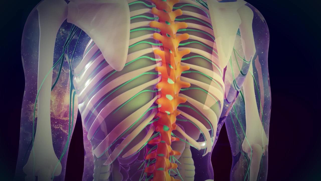
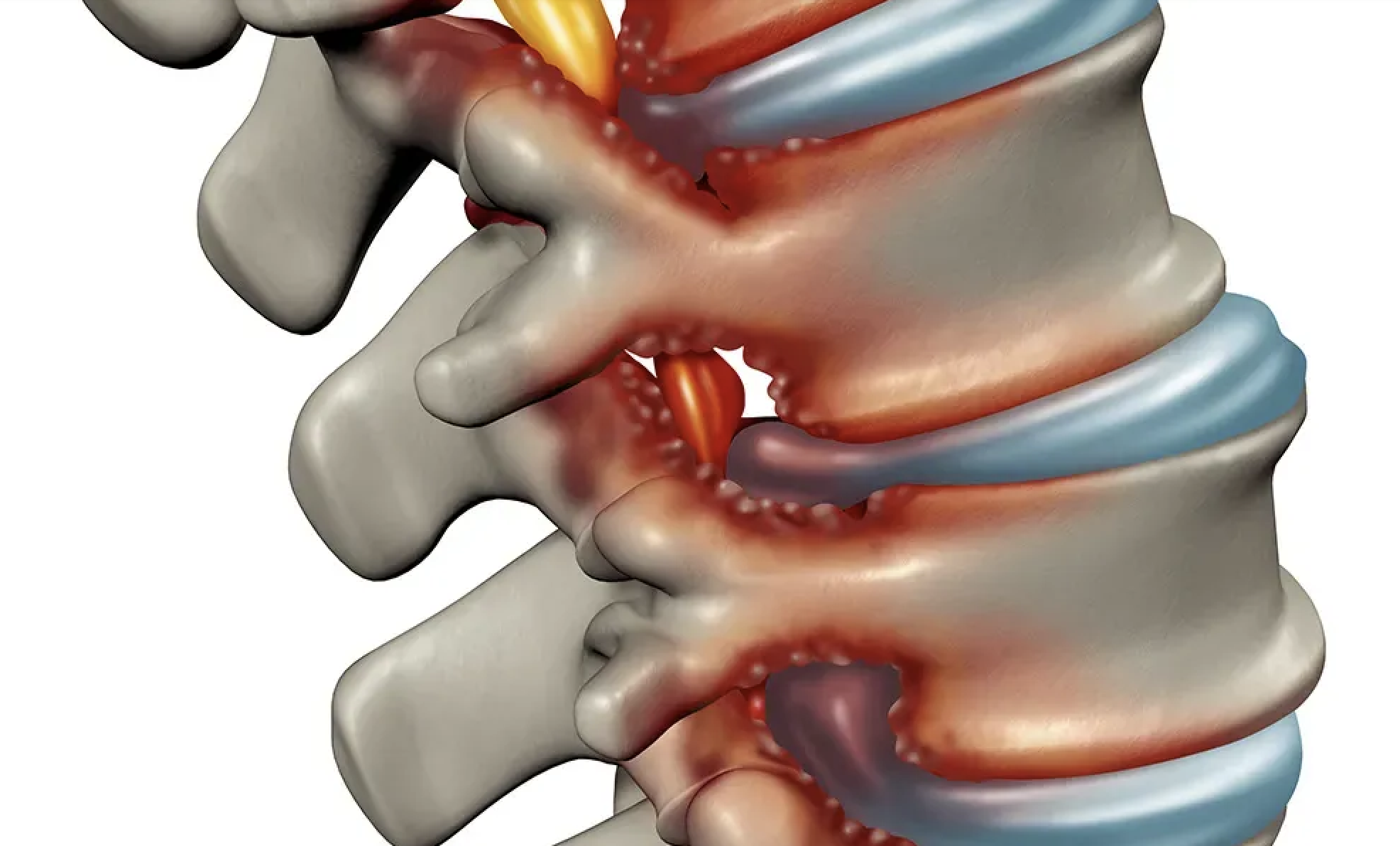
Herniated discs can compress nearby nerve roots or the spinal cord, leading to serious symptoms such as muscle weakness, loss of sensation, or mobility issues. In severe cases, this compression can result in partial paralysis or urinary incontinence, necessitating urgent surgical intervention.
Spinal instability
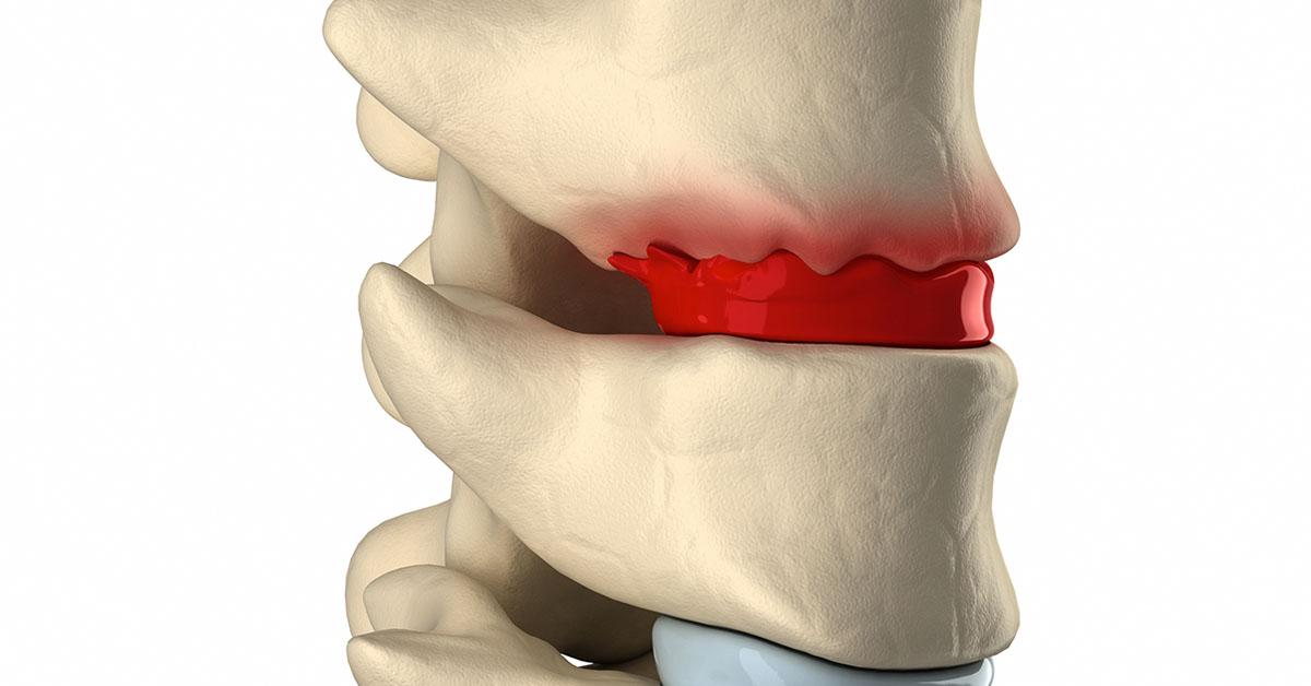
If a herniated disc is accompanied by spinal instability, spinal fusion may be recommended. Instability can cause abnormal movement between vertebrae, worsening pain and increasing the risk of complications.
Recurrence of a herniated disc
In some instances, a herniated disc may recur after initial surgical removal, either at the same site or in another segment of the spine. If this leads to severe symptoms, spinal fusion may be required to provide long-term stability to the spine.
Benefits of spinal fusion for herniated discs
Spinal fusion offers several advantages for patients with severe or complex herniated discs. These include:
- Long-lasting pain relief: By stabilizing the spine, spinal fusion reduces nerve compression, which is often the source of radiating pain.
- Improved mobility: Although the procedure limits movement at the fused vertebrae, it helps patients regain overall functional ability and quality of life.
- Reduced risk of recurrence: Fusing the vertebrae minimizes the likelihood of new herniated discs forming at the same level.
How is spinal fusion performed?
Pre-surgery preparation
Before the surgery, patients undergo thorough imaging tests such as MRIs or CT scans to precisely locate the herniation and assess vertebral health.
The procedure
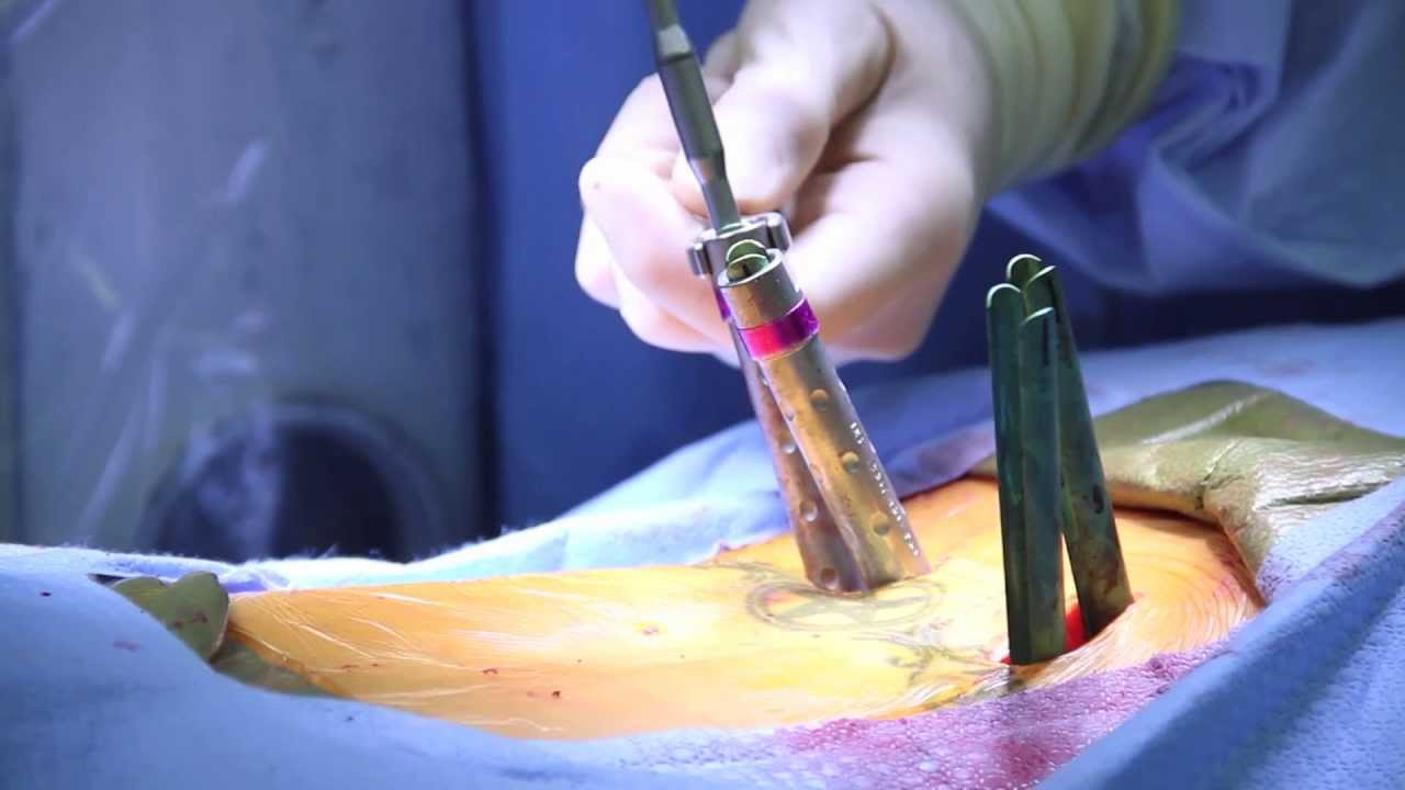
The surgery is performed under general anesthesia. The surgeon accesses the spine, removes the portion of the disc causing nerve compression, and places bone grafts and fixation devices to stabilize the vertebrae. The procedure’s duration varies depending on the complexity of the case.
Post-surgery recovery
Following the surgery, patients undergo a recovery period that includes physical therapy and rehabilitation. These steps are essential to restore mobility and strengthen the supporting muscles of the spine.
Is spinal fusion always the best option?
It’s important to note that spinal fusion is not the only solution for herniated discs. Every case is unique, and treatment decisions depend on factors such as the patient’s age, the overall condition of their spine, and the severity of symptoms. Other procedures, like discectomy or endoscopic techniques, may be sufficient in some cases.
A specialist’s evaluation is critical to determine the most appropriate course of action. A comprehensive assessment ensures the chosen treatment aligns with the patient’s needs, balancing the risks and benefits.
Your consultation with a spine specialist
Spinal fusion is a highly effective solution for patients with complex herniated discs or associated spinal instability. While it is typically a last resort after conservative treatments have failed, it offers significant, long-term relief from pain and improvements in quality of life.
If you have questions about this procedure or wish to assess your condition, you can schedule a consultation with a specialist. Virtual consultations are available for international patients seeking expert advice. By exploring your options, you can make informed decisions about your care and take the first step toward lasting relief.


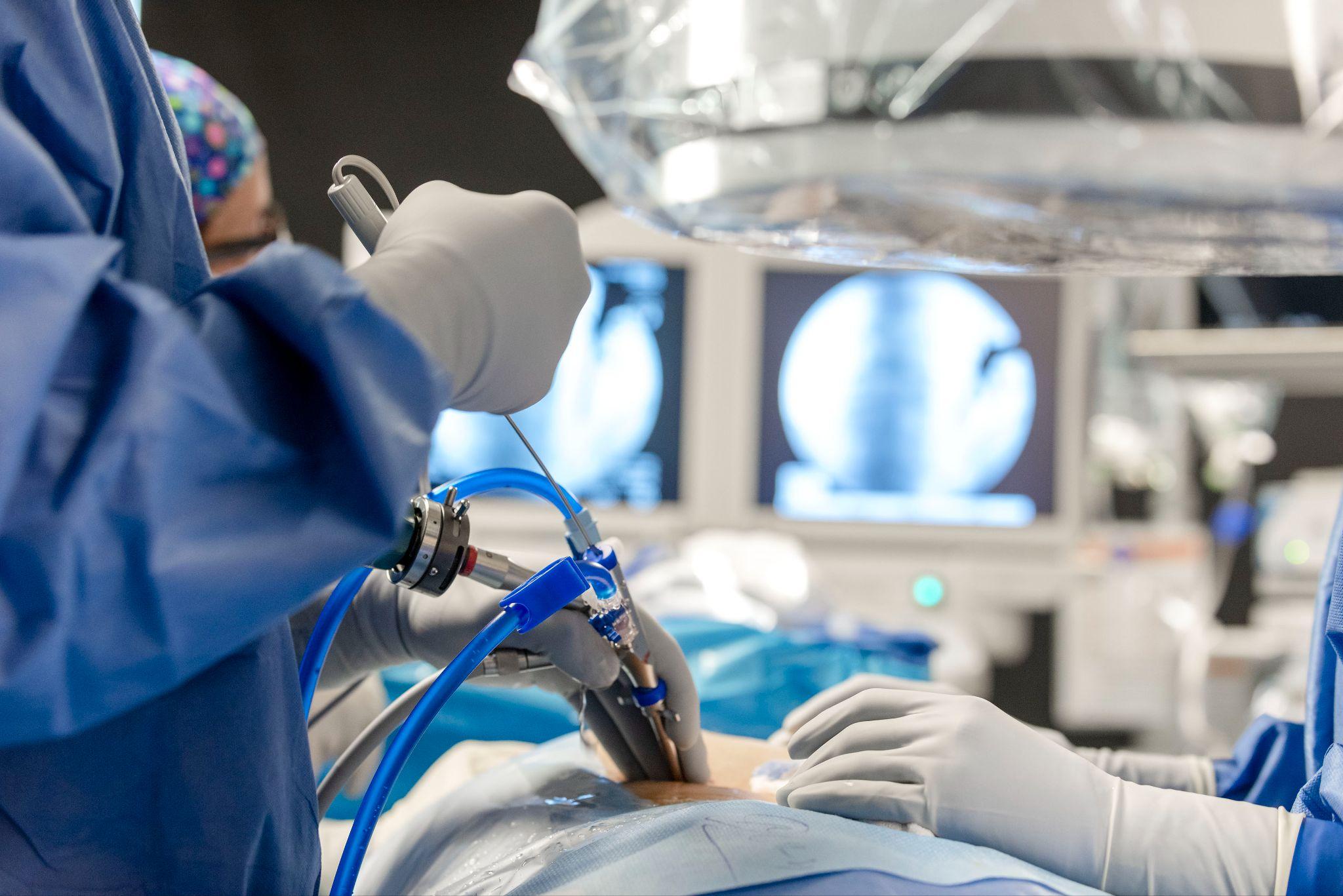
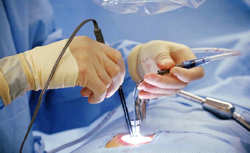
 Professionnal sportive could be operated by minimal invasive technique